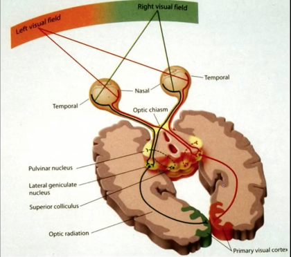
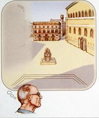
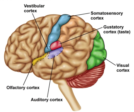
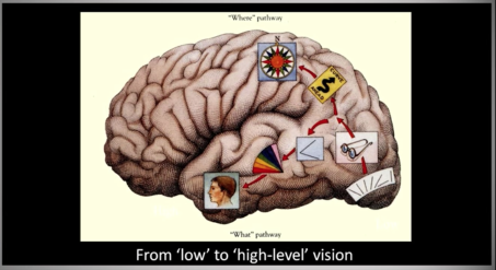
Visual Field
Monocular Visual Field: each eye $160^\circ$ (h)
Binocular Visual Field: $120^\circ$ (h)
Total Visual Field: $200^\circ$ (h)x$135^\circ$ (v)


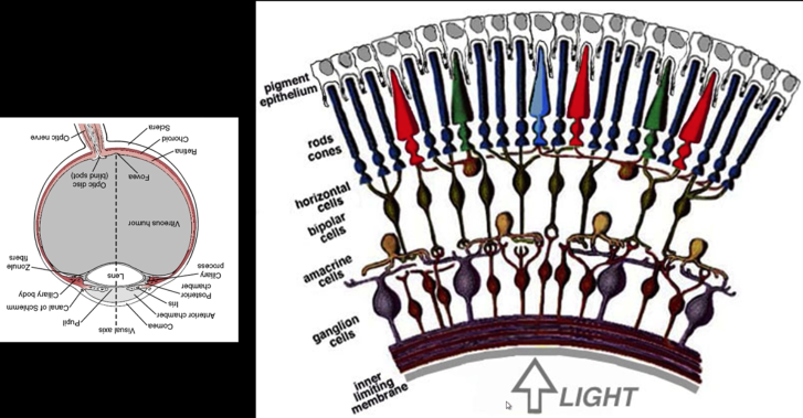
Rods and Cones

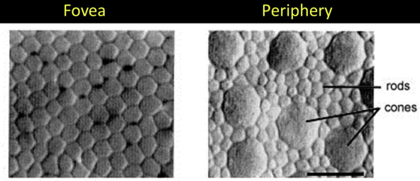
Rods:
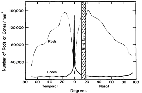
- 120 million rods in the retina
- 1000X more light sensitive than cones
- Discriminate between brightness levels, in low illumination
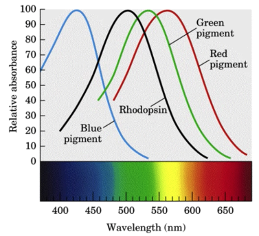
- Short-wavelength sensitive
Cones:
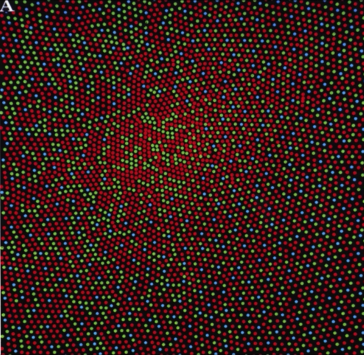
- 6-7 million cones in the retina
- Responsible for high-resolution vision
- Discriminate $\color{red}{c}\color{maroon}{o}\color{blue}{l}\color{green}{o}\color{pink}{r}\color{yellow}{s}$
- There are three types of color sensors: $64\%\,\color{red}{\text{ red, }}32\%\color{green}{\text{ green, }}2\%\color{blue}{\text{ blue}}$
- Sensitive to any combination of the three
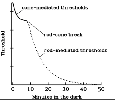


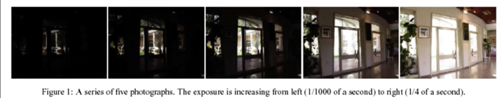
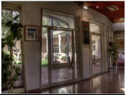
Dynamic Range
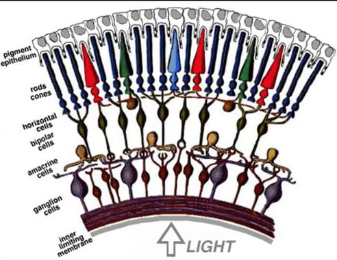
Image capture
- Huge dynamic range
- Overall: $10^{-6}$-$10^{+8}$ cd/$m^{2}$ (candelas)
- Static: at least 100:1 probably more
- A given scene in the real world: 100,000:1
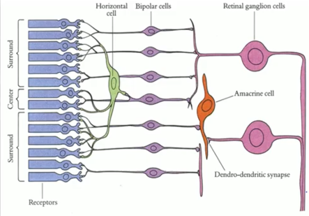
- Everyone knows about the pupil, but it's actually the retina's ganglion cells that make this works


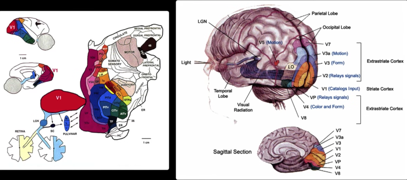


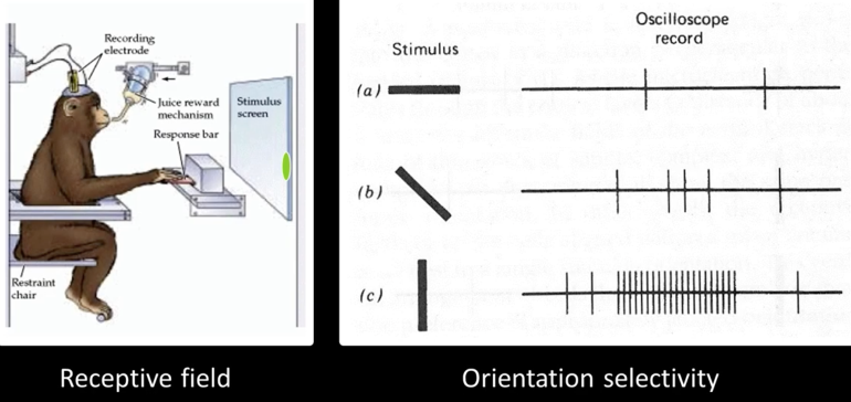
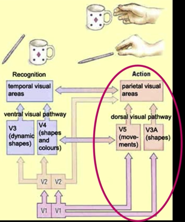
V2 and IT
Hunting for features in V2
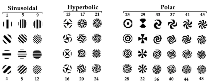
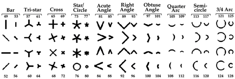
Increasing complexity
Inferotemporal cortex Features
K. Tanka, Neuronal Mechanisms of object Recognition Science, 1993

'Hand neuron' in area IT
Desimone, Albright, Gross and Bruce
Stimulus-selective properties of inferior temporal neurons in the macaque J Neurosci. 1984
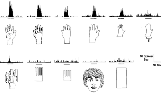
Some images look somewhat similar but represent different things
These fire similar cells in V1 but different cells in IT

Other images look very different but are the same thing. These fire very different cells in V1 but the same cells in inferior temporal cortex
fMRI and Facial Response
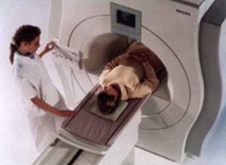
fMRI Magnet
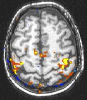
fMRI Activation

fMRI Activation Slice
FFA

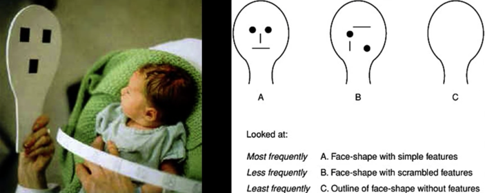
Faces are special: Early preference for faces?
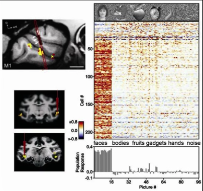
Face-selective Responses

All cells in this small area respond to faces
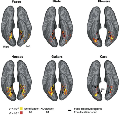
And more object specific areas
More Pathways

MT motion blindness
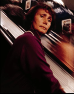
Gisela Leibold -- Unable to see motion, feels anxious as she rides down an escalator in Munich
She could not cross a street, because the motion of cars was invisible to her: a car was up the streen and then upon here, without ever seeming to occupy the intervening space.